Ankle
Proximally to the ankle joint, in the distal portions of the fibula and tibia, we find an important joint: the distal tibiofibular joint (syndemosis). It is formed by the rough, convex surface of the medial surface of the distal end of the fibula and a rough, concave surface of the lateral surface of the tibia. This joint is formed by the anterior and posterior tibiofibular, inferior transverse and interosseous ligaments.
Anterior Tibiofibular Ligament – It is a flattened fiber bundle that extends obliquely, distally, and laterally between the adjacent edges of the tibia and fibula, on the anterior surface of the syndesmosis.
Posterior Tibiofibular Ligament – Smaller than the anterior, it is arranged similarly to the posterior surface of the syndesmosis.
Inferior Transverse Ligament – It lies anterior to the posterior ligament and is a thick and robust bundle of yellowish fibers that run transversely from the lateral malleolus to the posterior edge of the articular surface of the tibia.
Interosseous Ligament – Consists of numerous fibrous bundles, short and robust, that pass between the continuous rough surface of the tibia and fibula and constitute the main bond of union between the bones.
The ankle joint proper is a hinge (hinge) formed by the distal end of the tibia and fibula. inferior transverse ligament and the talus.
The bones are connected by the joint capsule and the following ligaments: deltoid, anterior talofibular, posterior talofibular, and calcaneofibular.
Articular Capsule – Covers the joint. The anterior part of the capsule is a wide and thin membranous layer. The posterior part of the capsule is very thin and formed mainly by transverse fibers.
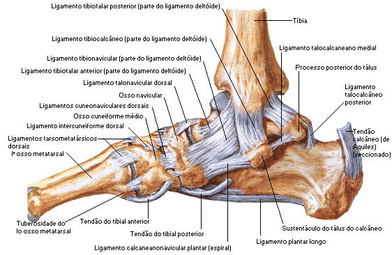
Deltoid Ligament – It is a triangular, robust and flattened bundle. It consists of two fiber bundles: superficial (tibionavicular, calcaneotibial and posterior talotibial fibers) and deep (anterior talotibial fibers). Its main function is to stabilize the medial region of the ankle and prevent the eversion movement.
- Tibionavicular Fibers – These are attached to the tuberosity of the navicular bone and posterior to this they unite with the medial border of the plantar calcaneavicular ligament.
- Fibers Calcaneotibial Fibers – They descend almost perpendicularly to insert along the entire length of the calcaneal talus support.
- Posterior Talotibial Fibers – Run laterally to attach to the inner side of the talus and the prominent tubercle on its posterior surface, medial to the groove for the flexor hallucis longus tendon.
- Anterior Talotibial Fibers – These are attached to the tip of the medial malleolus and the medial aspect of the talus.
Anterior Talofibular Ligament – Runs anteriorly and medially from the anterior border of the peroneal malleolus to the talus, anterior to its lateral articular facet.
Posterior Talofibular Ligament – Runs almost horizontally from the depression in the medial and posterior part of the peroneal malleolus to a prominent tubercle on the posterior aspect of the talus, just lateral to the groove for the flexor hallucis longus tendon.
Calcaneofibular Ligament – It is a narrow, rounded cord that runs from the apex of the fibular malleolus to a tubercle on the lateral surface of the calcaneus.
The three ligaments described above are collaterally referred to as the Lateral Collateral Ligament . It supports the lateral aspect of the ankle, preventing inversion movement.
The anterior and calcaneofibular ligaments are the most frequently injured in inversion ankle sprains. This is because with the foot in plantar flexion, the talus is more unstable in the ankle socket, and therefore more dependent on ligament support.
Distally to the joints studied above, we also find the intertarsal, tarsometatarsal, intermetatarsal, metatarsophalangeal and finger joints.
| MEDIAL VIEW OF ANKLE JOINT STRUCTURES |
 |
| Source: NETTER, Frank H.. Atlas of Human Anatomy. 2nd edition Porto Alegre: Artmed, 2000. |
| SIDE VIEW OF ANKLE JOINT STRUCTURES |
 |
| Source: NETTER, Frank H.. Atlas of Human Anatomy. 2nd edition Porto Alegre: Artmed, 2000. |
