occipital bone
It is pierced by a large, oval opening, the foramen magnum, through which the cranial cavity communicates with the vertebral canal. It has two portions: scaly and basilar.
a) Squamous – curved blade that extends posterior to the occipital foramen.
b) Basilar – anterior to the occipital foramen and thick.
scaly:
External face : posterior and convex. It has the following structures:
- External Occipital Protuberance – located between the apex of the bone and the foramen magnum
- External Occipital Crest
- Supreme Occipital (Nucal) Line – site of insertion of the galea aponeurotic. The external occipital protuberance is located laterally
- Superior Occipital (Nuchal) Line – located below the superior nuchal line
- Inferior Occipital (Nuchal) Line – just below the superior nuchal line
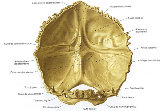
Inner Face : located anteriorly. It has the following structures:
- Cruciform Eminence – divides the inner face into four fossae
- Internal Occipital Protuberance – point of intersection of the four divisions
- Sagittal sulcus – houses the posterior portion of the superior sagittal sinus
- Internal Occipital Crest – inferior portion of the cruciform eminence
- Transverse Sinus Groove – lateral to the internal occipital bulge
- Superior Occipital Fossa (Brain)
- Inferior Occipital Fossa (Cerebellar)
Basic :
- foramen magnum – large oval opening that gives passage to the medulla oblongata (brain stem – medulla) and its membranes (meninges), cerebrospinal fluid, nerves, arteries, veins and ligaments.
Side :
- Occipital Condyles – are oval in shape and articulate with the 1st cervical vertebra (Atlas)
- Hypoglossal Canal – small excavation at the base of the occipital condyle that gives exit to the hypoglossal nerve (12th cranial nerve) and entry to a meningeal branch of the ascending pharyngeal artery.
- Condylar canal – next to the foramen magnum (gives the way to veins)
- Jugular Process – located lateral to the occipital condyle
The Occipital articulates with six bones: Parietal (2), Temporal (2), Sphenoid and Atlas.
| OCCIPITAL - EXTERNAL VIEW |
 |
| Source: SOBOTTA, Johannes. Atlas of Human Anatomy. 21 ed. Rio de Janeiro: Guanabara Koogan, 2000. |
| OCCIPITAL - INTERNAL VIEW |
 |
| Source: SOBOTTA, Johannes. Atlas of Human Anatomy. 21 ed. Rio de Janeiro: Guanabara Koogan, 2000 . |
