Arterial System
Set of vessels that leave the heart and branch successively, distributing themselves throughout the body. From the heart come the pulmonary trunk (it is related to the small circulation, that is, it takes venous blood to the lungs through its branch, two pulmonary arteries, one right and one left) and the aorta artery (carries arterial blood to the whole body through of its branches).
Some important arteries of the human body:
1 – Pulmonary Trunk System : the pulmonary trunk leaves the heart through the right ventricle and  bifurcates into two pulmonary arteries, one right and one left. Each of these branches from the pulmonary hilum into segmental pulmonary arteries.
Upon entering the lungs, these branches divide and subdivide to form capillaries around alveoli in the lungs. Carbon dioxide passes from the blood into the air and is exhaled. Oxygen passes from the air inside the lungs to the blood. This mechanism is called HEMATOSIS .
2 – Aorta Artery System (oxygenated blood): It is the largest artery in the body, with a diameter of 2 to 3 cm. Its four main divisions are the ascending aorta, the arch of the aorta, the thoracic aorta, and the abdominal aorta. The aorta is the main trunk of the systemic arteries. The part of the aorta that emerges from the left ventricle, posterior to the pulmonary trunk, is the ascending aorta.

The beginning of the aorta contains the aortic semilunar valves.
The aorta artery branches in the ascending portion into two coronary arteries, one right and one left that will supply the heart.
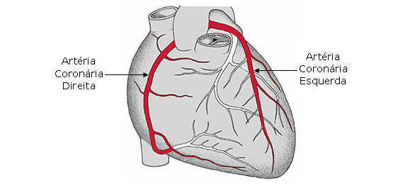
The Left Coronary Artery passes between the left atrium and the pulmonary trunk. It divides into two branches: anterior interventricular branch (left anterior descending branch) and a circumflex branch. The anterior interventricular branch passes along the interventricular sulcus toward the apex of the heart and supplies both ventricles. The circumflex branch follows the coronary sulcus around the left margin to the posterior surface of the heart, thus giving rise to the left marginal artery that supplies the left ventricle.
The Right Coronary Artery runs in the coronary or atrioventricular sulcus and gives rise to the right marginal branch that supplies the right margin of the heart as it runs to the apex of the heart. After originating these branches, it curves to the left and continues the coronary sulcus to the posterior surface of the heart, then emits the large posterior interventricular artery that descends in the posterior interventricular sulcus towards the apex of the heart, supplying both ventricles.
| CORONARY ARTERIES |
 |
| Source: NETTER, Frank H.. Atlas of Human Anatomy. 2nd edition Porto Alegre: Artmed, 2000. |
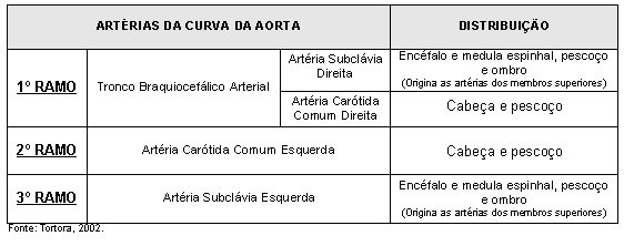
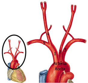 Soon after, the aorta artery curves forming an arc to the left, giving rise to three arteries (arteria of the aortic curve) which are:
1 – Arterial Brachiocephalic Trunk
2 - Left common carotid artery
3 - Left Subclavian Artery
The arterial brachiocephalic trunk gives rise to two arteries:
4 - Right Common Carotid Artery
5 - Right Subclavian Artery
ARTERIES OF THE NECK AND HEAD
The right and left vertebral arteries and the right and left common carotid arteries are responsible for the arterial vascularization of the neck and head.
Before entering the axilla, the subclavian artery gives a branch to the brain, called the vertebral artery, which passes through the transverse foramina from C6 to C1 and enters the skull through the foramen magnum. The vertebral arteries unite to form the basilar artery (supplies the cerebellum, pons and inner ear), which will give rise to the posterior cerebral arteries, which supply the inferior and posterior surface of the brain.
 At the upper border of the larynx, the common carotid arteries divide into the external carotid artery and the internal carotid artery.
The external carotid artery supplies the external structures of the skull. The internal carotid artery enters the skull through the carotid canal and supplies its internal structures. The terminal branches of the internal carotid artery are the anterior cerebral artery (supplies most of the medial surface of the brain) and middle cerebral artery (supplies most of the lateral surface of the brain).
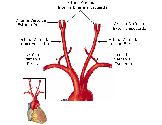
External carotid artery: supplies neck and face. Its collateral branches are: superior thyroid artery, lingual artery, facial artery, occipital artery, posterior auricular artery and ascending pharyngeal artery. Its terminal branches are: temporal artery and maxillary artery.
Willis Polygon :
Cerebral vascularization is formed by the right and left vertebral arteries and the right and left internal carotid arteries.
The vertebral arteries are anastomosed to originate the basilar artery, housed in the basilar gutter, it divides into two posterior cerebral arteries that supply the posterior part of the inferior surface of each of the cerebral hemispheres.
The internal carotid arteries on each side give rise to a middle cerebral artery and an anterior cerebral artery.
The anterior cerebral arteries communicate through a branch between them which is the anterior communicating artery.
The posterior cerebral arteries communicate with the internal carotid arteries through the posterior communicating arteries.
To learn more about the Polygon of Willis, see Nervous System (Vascularization of the Brain).
| WILLIS POLYGON |
 |
| Source: NETTER, Frank H.. Atlas of Human Anatomy. 2nd edition Porto Alegre: Artmed, 2000. |
| WILLIS POLYGON - SCHEME |
 |
| Source: NETTER, Frank H.. Atlas of Human Anatomy. 2nd edition Porto Alegre: Artmed, 2000. |
ARTERIES OF UPPER MEMBERS
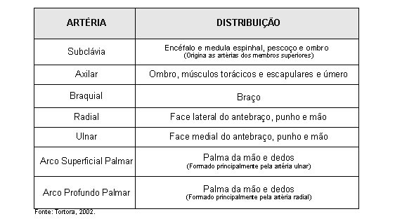
Explanation of the table above: the subclavian artery (right or left), soon after its beginning, gives rise to the vertebral artery that will assist in cerebral vascularization, descending towards the axilla is called the axillary artery, and when it finally reaches the arm , its name changes to brachial (humeral) artery. In the elbow region, it emits two terminal branches that are the radial and ulnar arteries that will travel through the forearm. In the hand, these two arteries anastomose forming a deep palmar arch that gives rise to the common palmar digital arteries and the palmar metacarpal arteries that will anastomose.

| ARTERIES OF THE UPPER LIMB |
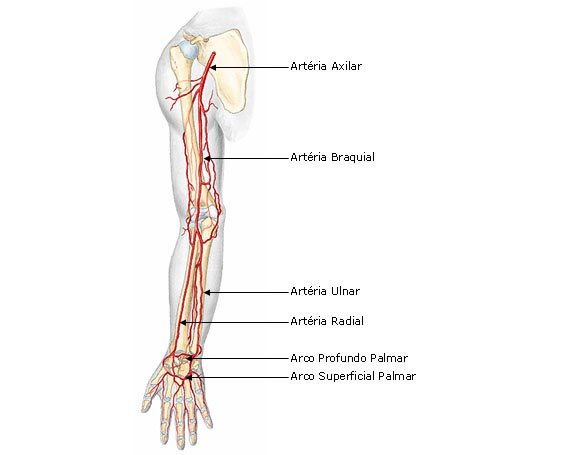 |
| Source: SOBOTTA, Johannes. Atlas of Human Anatomy. 21 ed. Rio de Janeiro: Guanabara Koogan, 2000. |
Aorta Artery – Thoracic Portion :
After the aotic curve or arch, the artery begins to descend from the left side of the spine, giving rise to the branches:
Visceral (nourish the organs):
1- Pericardial
2- Bronchial
3- Esophageal
4- Mediastinal
Parietals (irrigate the wall of the organs):
5- Posterior intercostals
6- Subcostals
7- Upper phrenics
Aorta Artery – Abdominal Portion :
When crossing the aortic hiatus of the diaphragm to the height of the fourth lumbar vertebra, where it ends, the aorta is represented by the abdominal portion.
In this portion the aorta provides several collateral branches and two terminals.
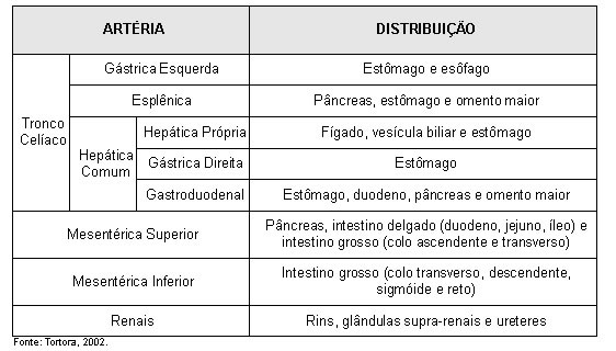
The terminal branches of the aorta artery are the right common iliac artery and the left common iliac artery.
| ARTERIES OF THE ABDOMINAL PORTION OF THE AORTA |
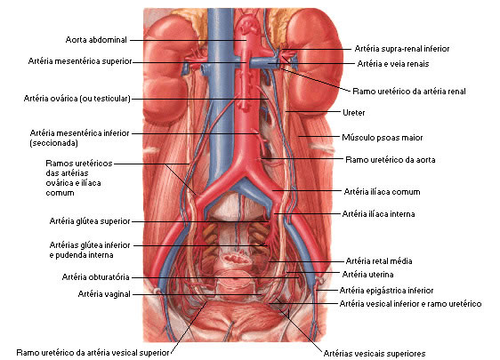 |
| Source: NETTER, Frank H.. Atlas of Human Anatomy. 2nd edition Porto Alegre: Artmed, 2000. |
| CELIAC TRUNK |
 |
| Source: NETTER, Frank H.. Atlas of Human Anatomy. 2nd edition Porto Alegre: Artmed, 2000. |
| BRANCHES OF THE SUPERIOR MESENTERIC ARTERY |
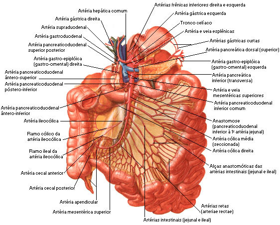 |
| Source: NETTER, Frank H.. Atlas of Human Anatomy. 2nd edition Porto Alegre: Artmed, 2000. |
| BRANCHES OF THE INFERIOR MESENTERIC ARTERY |
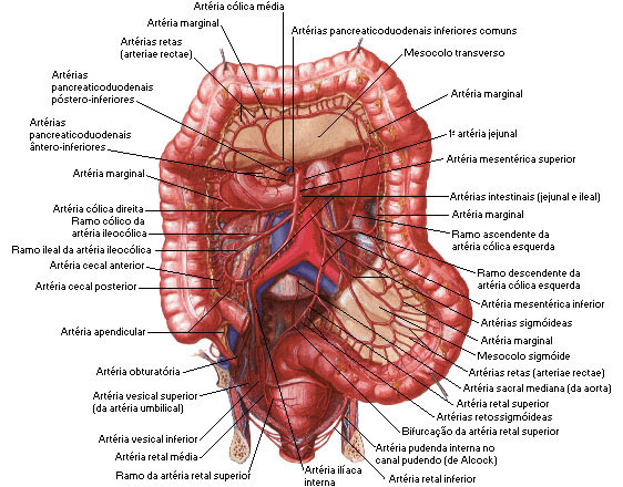 |
| Source: NETTER, Frank H.. Atlas of Human Anatomy. 2nd edition Porto Alegre: Artmed, 2000. |
| MAIN BRANCHES OF THE MESENTERIC ARTERIES |
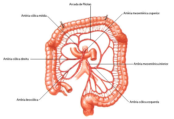 |
| Source: NETTER, Frank H.. Atlas of Human Anatomy. 2nd edition Porto Alegre: Artmed, 2000. |
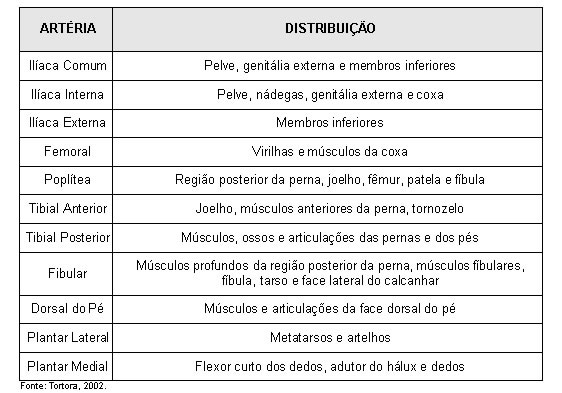
ARTERIES OF THE LOWER LIMBS
 |
| LOWER LIMBS ARTERIES |
 |
| Source: SOBOTTA, Johannes. Atlas of Human Anatomy. 21 ed. Rio de Janeiro: Guanabara Koogan, 2000. |


