Cerebellum
The cerebellum, an organ of the suprasegmental nervous system, derives from the dorsal part of the metencephalon and is located dorsal to the medulla and pons, contributing to the formation of the roof of the fourth ventricle. It rests on the cerebellar fossa of the occipital bone and is separated from the occipital lobe by a fold of dura mater called the tent of the cerebellum.
It is connected to the medulla and medulla by the inferior cerebellar peduncle and to the pons and midbrain by the middle and superior cerebellar peduncles, respectively. From a physiological point of view, the cerebellum fundamentally differs from the brain because it always functions at an involuntary and unconscious level, its function being exclusively motor (balance and coordination).
Anatomically 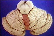 , distinguishes in the cerebellum, an odd and median portion, the vermis, connected to two large lateral masses, the cerebellar hemispheres. The vermis is slightly separated from the hemispheres on the upper surface of the cerebellum, which is not the case on the lower surface, where two very evident grooves separate it from the lateral parts.
| CEREBELLUM - LOWER VIEW |
 |
| Source: NETTER, Frank H.. Atlas of Human Anatomy. 2nd edition Porto Alegre: Artmed, 2000. |
| CEREBELLUM - TOP VIEW |
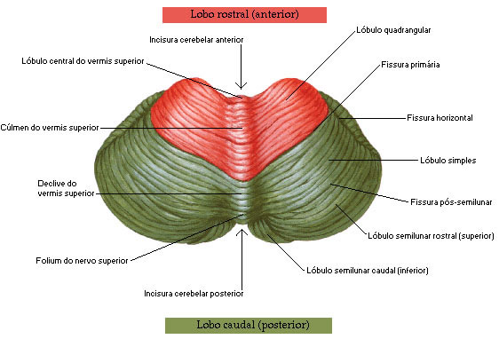 |
Source: NETTER, Frank H.. Atlas of Human Anatomy. 2nd edition Porto Alegre: Artmed, 2000. |
The surface presents grooves in a predominantly transverse direction, which delimit thin sheets called cerebellar leaves. There are also more pronounced grooves, the fissures of the cerebellum, which delimit lobes, each of which may contain several leaves. This arrangement, visible on the surface of the cerebellum, is especially evident in sections of this organ, which also give an idea of its internal organization. It is thus seen that the cerebellum is made up of a center of white matter, the medullary body of the cerebellum, from which radiates the white lamina of the cerebellum, covered externally by a thin layer of gray matter, the cerebellar cortex. The medullary body of the cerebellum with its white blades, when seen in sagittal sections, is called the “tree of life”. Within the medullary field there are four pairs of gray matter nuclei, which are the central nuclei of the cerebellum: dentate, emboliform, globose and fastigial.
| CEREBELLUM - SECTION IN THE PLAN OF THE SUPERIOR CEREBELAR PEDUNCLE |
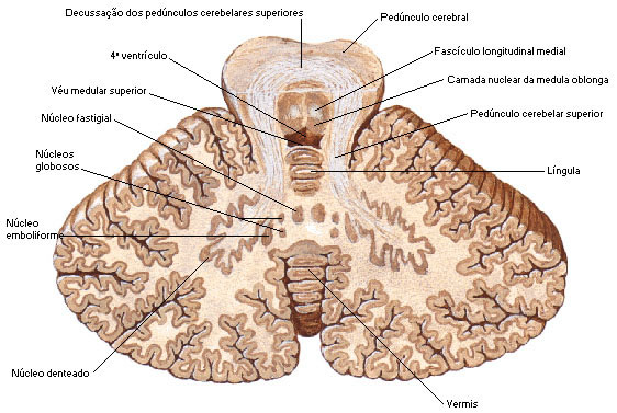 |
| Source: NETTER, Frank H.. Atlas of Human Anatomy. 2nd edition Porto Alegre: Artmed, 2000. |
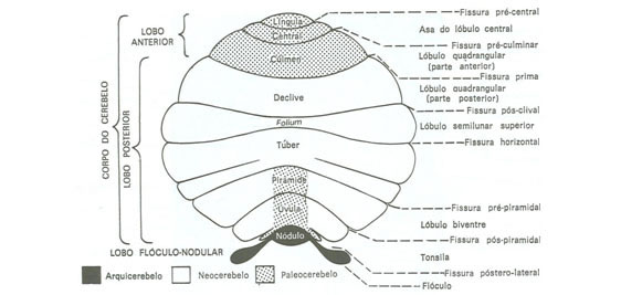
Cerebellar lobes : the division of the cerebellum into lobules has no functional significance and its importance is only topographical. The lobes receive different denominations in the vermis and in the hemispheres. Each lobe of the vermix corresponds to two hemispheres.
The lingula is almost always attached to the superior medullary veil. The folium consists of just one leaf of the vermix. An important lobe is the flocculus, situated just below the point where the middle cerebellar peduncle enters the cerebellum, close to the vestibulocochlear nerve. It is connected to the nodule, lobe of the vermix, by the peduncle of the floccule. The tonsils are quite evident in the lower part of the cerebellum, projecting medially over the dorsal surface of the medulla.
| CEREBELLUM - MEDIAL VIEW |
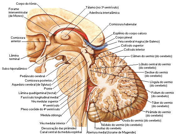 |
| Source: NETTER, Frank H.. Atlas of Human Anatomy. 2nd edition Porto Alegre: Artmed, 2000. |
Cerebellar fissures :
– After the lingula we have the precentral fissure .
– After the central lobe we have the pre-culminating fissure .
– After the culmen we have the prima fissure .
– After the slope we have the post-clival fissure .
– After the folium we have the horizontal fissure .
– After the tuber we have the pre-pyramidal fissure .
– After the pyramid we have the post-pyramidal fissure .
– After the uvula we have the posterolateral fissure .

| CEREBELLUM - SIDE VIEW |
 |
| Source: MACHADO, Ângelo. Functional Neuroanatomy. Rio de Janeiro/São Paulo: Atheneu, 1991. |
