Digestive system
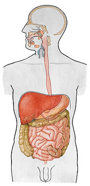
The digestive tract and associated organs make up the digestive system. The digestive tract is a hollow tube that extends from the oral cavity to the anus, being also called the alimentary canal or gastrointestinal tract. The structures of the digestive tract include: Mouth, Pharynx, Esophagus, Stomach, Small Intestine, Large Intestine, Rectum and Anus.
FUNCTIONS:
2- Mechanical and chemical transformation of ingested food macromolecules (proteins, carbohydrates, etc.) into molecules of suitable sizes and shapes to be absorbed by the intestine.
3- Transport of digested food, water and mineral salts from the intestinal lumen to the blood capillaries of the intestinal mucosa.
4- Elimination of undigested and unabsorbed food residues together with the remains of cells desquamated from the part of the gastro intestinal tract and substances secreted in the lumen of the intestine.
Chewing: Partial disintegration of food, mechanical and chemical process.
Swallowing : Conduction of food through the pharynx into the esophagus.
Ingestion : Introduction of food into the stomach.
Digestion : Breaking down food into simpler molecules.
Absorption : Process carried out by the intestines.
Defecation : Elimination of undigested substances from the gastrointestinal tract.
The gastrointestinal tract has several segments which are successively: MOUTH, PHARYNX, ESOPHAGUS, STOMACH, SMALL INTESTINE, LARGE INTESTINE, RECTUM and ANUS.
Attached Bodies :
- PAROTID GLANDS
- SUBMANDIBULAR GLANDS
- SUBLINGUAL GLANDS
- LIVER
- PANCREAS

MOUTH
The mouth, also referred to as the Oral or Buccal Cavity , is formed by the cheeks (they form the side walls of the face and are constituted externally by skin and internally by mucosa), by the hard (upper wall) and soft (posterior wall) palates and by the tongue (important for transporting food, sense of taste and speech). The soft palate extends posteriorly into the oral cavity as the uvula, which is a V-shaped structure that is suspended in the upper and posterior region of the oral cavity.
Limits of the Oral Cavity

| BUCCAL CAVITY |
 |
| Source: NETTER, Frank H.. Atlas of Human Anatomy. 2nd edition Porto Alegre: Artmed, 2000. |
| HARD AND SOFT PALATE |
 |
| Source: NETTER, Frank H.. Atlas of Human Anatomy. 2nd edition Porto Alegre: Artmed, 2000. |
| SOFT PALATE |
 |
| Source: NETTER, Frank H.. Atlas of Human Anatomy. 2nd edition Porto Alegre: Artmed, 2000. |
The mouth cavity is where food is ingested and prepared for digestion in the stomach and small intestine. Food is chewed by the teeth, and saliva, from the salivary glands, facilitates the formation of a controllable food bolus. Swallowing is initiated voluntarily in the mouth cavity. The voluntary phase of the process pushes the bolus from the mouth cavity into the pharynx – the expanded part of the digestive tract – where the automatic phase of swallowing takes place.
The mouth cavity consists of two parts: the vestibule of the mouth and the mouth cavity proper . The vestibule of the mouth is the cleft-like space between the teeth and gums and the lips and cheeks. The mouth cavity proper is the space between the upper and lower dental arches. It is limited laterally and anteriorly by the maxillary and mandibular alveolar arches that house the teeth. The roof of the mouth cavity is formed by the palate. Subsequently, the mouth cavity communicates with the oral part of the pharynx. When the mouth is closed and at rest, the mouth cavity is completely occupied by the tongue. 
Teeth
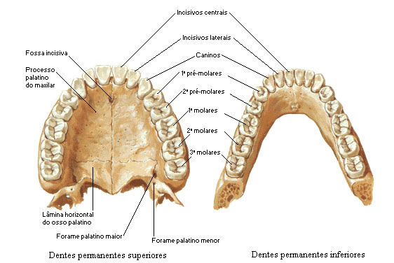
Teeth are hard, conical structures attached to the sockets of the mandible and maxilla that are used in mastication and speech assistance.
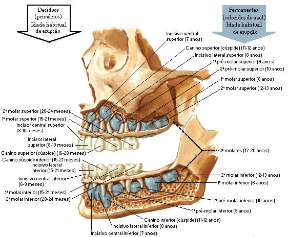
Children have 20 deciduous teeth (primary or milk). Adults normally have 32 secondary teeth. By the time your child is 2 years old, they will likely have a full set of 20 baby teeth. By the time a young adult is between the ages of 17 and 24, a full set of 32 permanent teeth is usually present in their mouth.
| PRIMARY AND PERMANENT TEETH |
 |
| Source: NETTER, Frank H.. Atlas of Human Anatomy. 2nd edition Porto Alegre: Artmed, 2000. |
| PERMANENT TEETH |
 |
| Source: NETTER, Frank H.. Atlas of Human Anatomy. 2nd edition Porto Alegre: Artmed, 2000. |
Tongue
The tongue is the main organ of the sense of taste and an important organ of speech, in addition to assisting in chewing and swallowing food. It is located on the floor of the mouth, within the curve of the body of the mandible.
The root is the posterior part, where it is connected to the hyoid bone by the hyoglossus and genioglossus muscles and by the glossioid membrane; to the epiglottis, by three folds of the mucosa; to the soft palate, by the palatoglossal arches, and the pharynx, by the superior pharyngeal constrictor muscles and the mucosa. 
The apex is the anterior, somewhat rounded end that rests against the lingual surface of the lower incisor teeth.

The lower surface has a mucosa between the floor of the mouth and the tongue in the midline that forms a sharp vertical fold, the lingual frenulum.
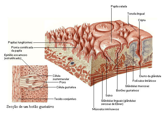
On the dorsum of the tongue we find a median groove that divides the tongue into symmetrical halves. On the anterior 2/3 of the dorsum of the tongue we find the lingual papillae. In the posterior 1/3 we find numerous mucous glands and lymphatic follicles (lingual tonsil).
Lingual papillae – are projections of the chorion, abundantly distributed in the anterior 2/3 of the tongue, giving this region a characteristic roughness. The types of papillae are: vallate, fungiform, filiform and simple papillae.
| TONGUE PAPILLS |
 |
| Source: NETTER, Frank H.. Atlas of Human Anatomy. 2nd edition Porto Alegre: Artmed, 2000. |
| TONGUE PAPILLS |
 |
Source: NETTER, Frank H.. Atlas of Human Anatomy. 2nd edition Porto Alegre: Artmed, 2000. |
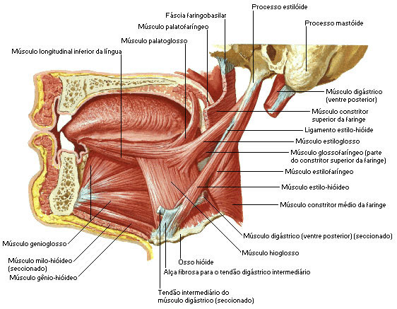
Muscles of the Tongue - The tongue is divided into halves by a median fibrous septum that runs its entire length and attaches inferiorly to the hyoid bone. In each half there are two sets of muscles, extrinsic and intrinsic.
The Extrinsic Muscles are: Genioglossus, Hyoglossus, Chondroglossus, Styloglossus and Palatoglossus.
The Intrinsic Muscles are: Superior Longitudinal, Inferior Longitudinal, Transverse and Vertical.
| TONGUE MUSCLES |
 |
| Source: NETTER, Frank H.. Atlas of Human Anatomy. 2nd edition Porto Alegre: Artmed, 2000. |
PHARYNX
The pharynx is a tube that extends from the mouth to the esophagus.
The pharynx has very thick walls due to the volume of the muscles that cover it externally, on the inside, the organ is lined by the pharyngeal mucosa, a smooth epithelium, which facilitates the rapid passage of food.
The movement of food from the mouth to the stomach is accomplished by the act of swallowing. Swallowing is facilitated by saliva and mucus and involves the mouth, pharynx and esophagus.
Three stages:
- Voluntary : in which the bolus is passed into the oral part of the pharynx.
- Pharyngeal : Involuntary passage of food through the pharynx into the esophagus.
- Esophageal : Involuntary passage of food through the esophagus into the stomach.
Pharyngeal Limits:
- Superior – body of sphenoid and basilar portion of occipital bone
- Lower – esophagus
- Posterior – spine and fascia of the long neck and long head muscles
- Anterior – pterygoid process, mandible, tongue, hyoid bone, and thyroid and cricoid cartilages
- Lateral – styloid process and its muscles
The pharynx can be further divided into three parts : nasal ( Nasopharynx ), oral ( Oropharynx ) and laryngeal ( Laryngopharynx ).
Nasal Part – It is located posterior to the nose and above the soft palate and differs from the other two parts in that its cavity is always open. It communicates anteriorly with the nasal cavities through the choanae. On the posterior wall is the pharyngeal tonsil (adenoid in children).
Oral part – extends from the soft palate to the hyoid bone. On its lateral wall is the palatine tonsil.
Laryngeal part – extends from the hyoid bone to the cricoid cartilage. On each side of the laryngeal orifice is a recess called the piriform sinus.
THE 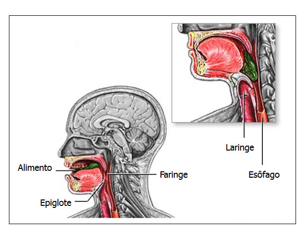 pharynx communicates with the nasal, respiratory and digestive tracts. The act of swallowing normally directs food from the throat into the esophagus, a long tube that empties into the stomach. During swallowing, food normally cannot enter the nasal and respiratory passages due to the temporary closure of the openings in these passages. Thus, during swallowing, the soft palate moves towards the opening of the nasal part of the pharynx; the opening of the larynx is closed as the trachea moves upward and allows a fold of tissue, called the epiglottis, to cover the entrance to the airway.
The movement of the larynx also simultaneously pulls the vocal cords and increases the opening between the laryngeal part of the pharynx and the esophagus. The food bolus passes through the laryngeal part of the pharynx and enters the esophagus in 1-2 seconds.
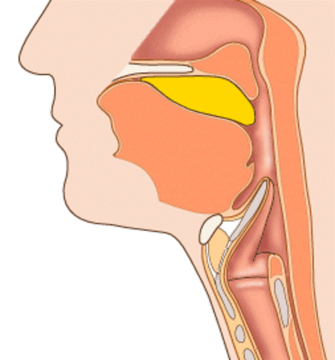
| PARTS AND STRUCTURE OF THE PHARYNX |
 |
| Source: NETTER, Frank H.. Atlas of Human Anatomy. 2nd edition Porto Alegre: Artmed, 2000. |
ESOPHAGUS
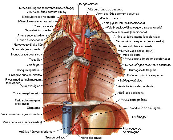
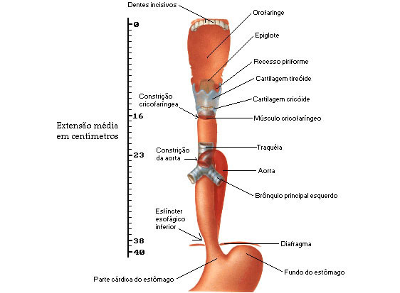
The esophagus is a fibro-musculo-mucous tube that runs between the pharynx and stomach. It is located posterior to the trachea starting at the level of the 7th cervical vertebra. It pierces the diaphragm through the opening called the esophageal hiatus and ends in the upper part of the stomach. It measures about 25 centimeters in length.
| PARTS AND STRUCTURE OF THE ESOPHAGUS |
 |
| Source: NETTER, Frank H.. Atlas of Human Anatomy. 2nd edition Porto Alegre: Artmed, 2000. |
The presence of food inside the esophagus stimulates peristaltic activity, and causes food to move into the stomach.
The contractions are repeated in waves that push the food toward the stomach. The passage of solid or semi-solid food from the mouth to the stomach takes 4 – 8 seconds; very soft foods and liquids pass about 1 second.
Occasionally, the reflux of stomach contents into the esophagus causes heartburn (or heartburn). The burning sensation is a result of the high acidity of the stomach contents.
Gastroesophageal reflux occurs when the lower esophageal sphincter (located at the top of the esophagus) does not close properly after food has entered the stomach, the contents can flow back down the esophagus.
The esophagus is made up of three parts:
- Cervical Portion : Portion that is in intimate contact with the trachea.
- Thoracic Portion : it is the most important portion, it passes behind the left bronchus (upper mediastinum, between the trachea and the vertebral column).
- Abdominal portion : rests on the diaphragm and presses on the liver, forming an esophageal impression on it.
| PARTS OF THE ESOPHAGUS |
 |
| Source: NETTER, Frank H.. Atlas of Human Anatomy. 2nd edition Porto Alegre: Artmed, 2000. |
STOMACH
The stomach is situated in the abdomen, just below the diaphragm, anterior to the pancreas, superior to the duodenum and to the left of the liver. It is partially covered by the ribs. The stomach is located in the upper left quadrant of the abdomen (See Abdominal quadrants in the main menu), between the liver and spleen.
The stomach is the most dilated segment of the digestive tract, because food remains in it for some time, it needs to be a reservoir between the esophagus and the small intestine.
The shape and position of the stomach varies greatly from person to person; the diaphragm pushes it down with each inhalation and pulls it up with each exhalation and so cannot be described as typical.
The stomach is divided into 4 main areas (regions): Cardia, Fundus, Body and Pylorus.
| PARTS AND STRUCTURE OF THE STOMACH |
 |
Source: NETTER, Frank H.. Atlas of Human Anatomy. 2nd edition Porto Alegre: Artmed, 2000. |
The fundus, which despite the name, is located at the top, above the point where the esophagus meets the stomach.
The body represents about 2/3 of the total volume.
To prevent the reflux of food into the esophagus, there is a valve (stomach inlet hole - cardiac ostium or lower esophageal orifice), the Cardia , located just above the lesser curvature of the stomach. It is so called because it is close to the heart.
To prevent the food bolus from passing into the small intestine prematurely, the stomach is equipped with a powerful muscular valve, a sphincter called the pylorus (stomach outlet - pyloric ostium).
Just before the pyloric valve we find a portion called antropyloric.
The stomach also has two parts: the Greater Curvature (left side of the stomach) and the Lesser Curvature (right side of the stomach). 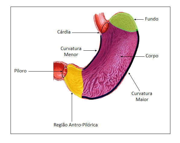
| PARTS OF THE STOMACH |
 |
| Source: NETTER, Frank H.. Atlas of Human Anatomy. 2nd edition Porto Alegre: Artmed, 2000. |
Digestive Functions of the Stomach:
- food digestion
- Secretion of gastric juice, which includes digestive enzymes and hydrochloric acid as the most important substances.
- Secretion of gastric hormone and intrinsic factor.
- Regulation of the pattern in which food is partially digested and delivered to the small intestine.
- Absorption of small amounts of water and dissolved substances.
SMALL INTESTINE
The main part of digestion takes place in the small intestine, which extends from the pylorus to the ileocolic (ileocecal) junction, which meets with the large intestine. The small intestine is an indispensable organ. The main events of digestion and absorption take place in the small intestine, so its structure is specially adapted for this function. Its length provides a large surface area for digestion and absorption, and is further increased by circular folds, villi and microvilli.
The small intestine removed in one is about 7 meters long, and can vary between 5 and 8 meters (the length of the small and large intestine together after death is 9 meters).
The small intestine, which consists of the duodenum, jejunum, and ileum , extends from the pylorus to the ileocecal junction where the ileum joins the cecum, the first part of the large intestine.
Duodenum : It is the first portion of the small intestine. It receives this name because its length is approximately equal to the width of twelve fingers (25 centimeters). It is the only portion of the small intestine that is fixed. It has no mesentery.
2) Descending part or 2nd portion – it is deperitonized and we find the arrival of two Ducts:
- Common bile duct – comes from the gallbladder and liver (bile)
- Pancreatic duct – comes from the pancreas (pancreatic juice or secretion)

| CHOLEDOCO DUCT AND ADJACENT STRUCTURES |
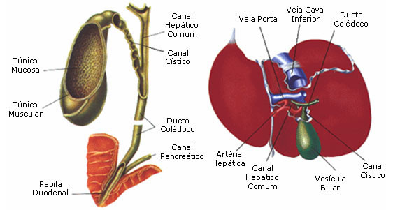 |
| CHOLEDOCO DUCT AND ADJACENT STRUCTURES |
 |
| Source: NETTER, Frank H.. Atlas of Human Anatomy. 2nd edition Porto Alegre: Artmed, 2000. |
3) Horizontal part or 3rd part
4) Ascending Part or 4th portion
Jejunum : it is the part of the small intestine that continues to the duodenum, it receives this name because whenever it is opened it is empty. It is wider (approximately 4 centimeters), its wall is thicker, more vascular and stronger in color than the ileum.
Ileum : It is the last segment of the small intestine that continues to the jejunum. It receives this name in relation to the iliac bone. It is narrower and its tunics are thinner and less vascularized than the jejunum. Distally, the ileum flows into the large intestine at an orifice called the ileocecal ostium.
Together, the jejunum and ileum measure 6 to 7 meters in length. Most of the jejunum is in the upper left quadrant, while most of the ileum is in the lower right quadrant. The jejunum and ileum, unlike the duodenum, are mobile.
| SMALL INTESTINE |
 |
| Source: NETTER, Frank H.. Atlas of Human Anatomy. 2nd edition Porto Alegre: Artmed, 2000. |
LARGE INTESTINE
The large intestine can be compared to a horseshoe, open downwards, measuring about 6.5 centimeters in diameter and 1.5 meters in length. It extends from the ileum to the anus and is attached to the posterior abdominal wall by the midneck.
The large intestine absorbs water so quickly that, in about 14 hours, the food material takes on the typical consistency of a fecal bolus.
The large intestine has some differences in relation to the small intestine: the caliber, the tapeworms, the haustos and the epiploic appendages.
The large intestine is larger than the small intestine, which is why it is called the large intestine. The caliber gradually gets thinner as it reaches the anal canal.
The tapeworms of the colon (longitudinal ribbons) are three bands approximately 1 centimeter wide that run along the entire length of the large intestine. They are most evident in the cecum and ascending colon.
Colonic hausts (sacculations) are ampullary bulges separated by transverse grooves.
The epiploic appendages are small yellowish pendants made up of connective tissue rich in fat. They appear mainly in the sigmoid colon.
The large intestine is divided into 4 main parts: Cecum (cecun), Colon (colun) (Ascending, Transverse, Descending and Sigmoid), Rectum and Anus.

The first is the cecum, the largest segment, which communicates with the ileum. To prevent the reflux of material from the small intestine, there is a valve located at the junction of the ileum with the cecum – Ileocecal (Ileocolic) Valve. At the bottom of the cecum, we find the Vermiform Appendix.
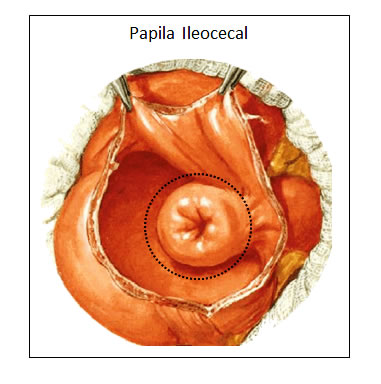
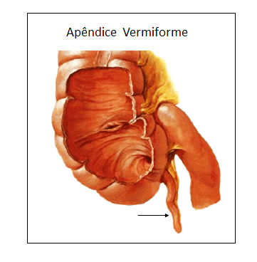
The next portion of the large intestine is the colon , a segment that extends from the cecum to the anus.
Ascending Colon – Transverse Colon – Descending Colon – Sigmoid Colon
| LARGE INTESTINE PARTS AND STRUCTURE |
 |
| Source: NETTER, Frank H.. Atlas of Human Anatomy. 2nd edition Porto Alegre: Artmed, 2000. |
Ascending Colon – is the second part of the large intestine. It passes upward on the right side of the abdomen from the cecum to the right lobe of the liver, where it curves to the left at the right flexure of the cervix (hepatic flexure).
Transverse colon – is the widest and most mobile part of the large intestine. It crosses the abdomen from the right flexure of the cervix to the left flexure of the cervix, where it curves inferiorly to become the descending colon. The left flexure of the cervix (splenic flexure), usually more superior, sharper, and less mobile than the right flexure of the cervix.
Descending Neck – passes retroperitoneally from the left flexure of the neck to the left iliac fossa, where it is continuous with the sigmoid colon.
Colon Sigmoid – is characterized by its handle in the shape of an “S”, of variable length. The sigmoid colon joins the descending colon to the rectum. The termination of the taenia coli, approximately 15 cm from the anus, indicates the recto-sigmoid junction.
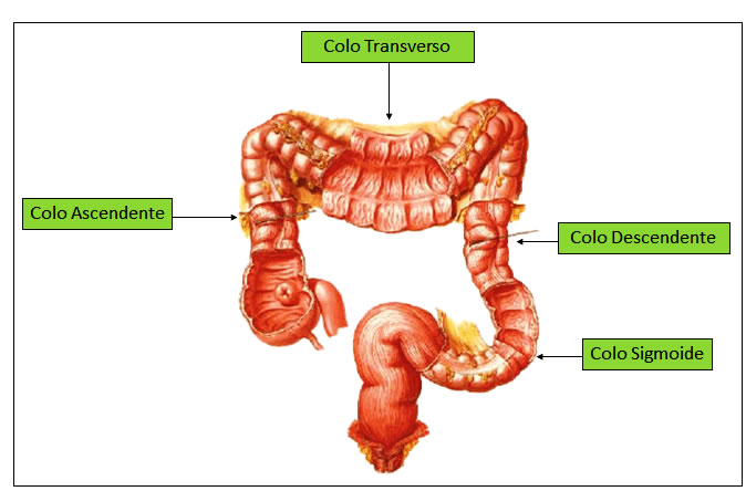
- Hepatic Flexure – between the ascending colon and the transverse colon.
- Splenic Flexure – between the transverse colon and the descending colon.

| LARGE INTESTINE DIVISIONS |
 |
| Source: NETTER, Frank H.. Atlas of Human Anatomy. 2nd edition Porto Alegre: Artmed, 2000. |
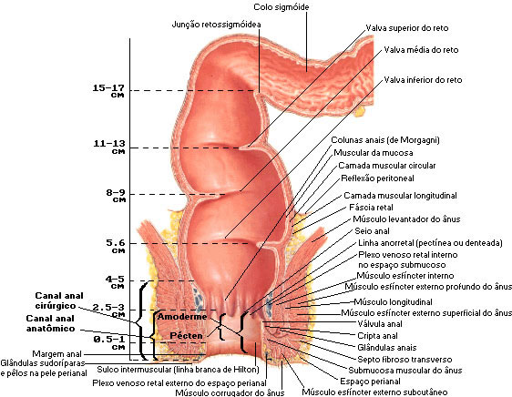
The rectum gets its name because it is almost straight. This segment of the large intestine ends when it pierces the pelvic diaphragm (the levator ani muscles) and becomes the anal canal.
The anal canal, despite being quite short (3 centimeters in length), is important because it presents some essential formations for intestinal functioning, of which we mention the anal sphincters.
The internal anal sphincter is the deepest, and results from a thickening of circular smooth muscle fibers, and is consequently involuntary. The external anal sphincter consists of striated muscle fibers that are arranged circularly around the internal anal sphincter, which is voluntary. Both sphincters must relax before defecation can occur.
| ANAL CANAL AND ANAL SPINCTER |
 |
| Source: NETTER, Frank H.. Atlas of Human Anatomy. 2nd edition Porto Alegre: Artmed, 2000 . |
Large Intestine Functions:
- Absorption of water and certain electrolytes;
- Synthesis of certain vitamins by intestinal bacteria;
- Temporary storage of waste (feces);
- Elimination of waste from the body (defecation).
Peristalsis:
Intermittent, well-spaced peristaltic waves move fecal material from the cecum into the ascending, transverse, and descending colon. As it moves through the colon, water is continually reabsorbed from the stool, through the walls of the intestine, into the capillaries. Stools that stay in the large intestine for a longer period of time lose excess water, developing so-called constipation. On the contrary, rapid bowel movements do not allow enough time for water reabsorption to occur, causing diarrhea.
PERITONEUM
The peritoneum is the most extensive serous membrane in the body. The part that lines the abdominal wall is called the Parietal Peritoneum and the part that reflects on the viscera constitutes the Visceral Peritoneum . The space between the parietal and visceral layers of the peritoneum is called the peritoneal cavity.
Certain abdominal viscera are completely surrounded by peritoneum and suspended from the wall by a thin sheet of serous-lined connective tissue containing the blood vessels. These folds are given the general name of mesentery.
The mesenteries are: the mesentery proper, the transverse mesocolon and the sigmoid mesocolon. In addition to these, an ascending and a descending mesocolon are sometimes present.
The Mesentery Proper – originates from the ventral structures of the spinal column and holds the small intestine suspended.
The Transverse Mesocolon – Attaches the transverse colon to the posterior wall of the abdomen.
The Sigmoid Mesocolon – maintains the sigmoid colon in connection with the pelvic wall.
The Ascending and Descending Mesocolon – connect the ascending and descending colon to the posterior abdominal wall.
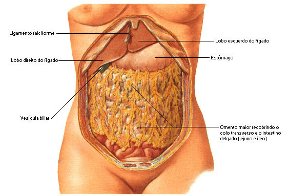
The peritoneum has two oments : the greater and the lesser.
The Greater Omentum is a thin apron that hangs over the transverse colon and the loops of the small intestine. It is inserted along the greater curvature of the stomach and the first portion of the duodenum.
The Lesser Omentum extends from the lesser curvature of the stomach and the initial portion of the duodenum to the liver.
Epiploic appendages – are small, fat-filled pouches of peritoneum located along the colon and upper part of the rectum.
| PERITONEUM STRUCTURES |
 |
| Source: NETTER, Frank H.. Atlas of Human Anatomy. 2nd edition Porto Alegre: Artmed, 2000. |
| MAJOR OMENTA |
 |
| Source: NETTER, Frank H.. Atlas of Human Anatomy. 2nd edition Porto Alegre: Artmed, 2000. |
ATTACHED BODIES
The digestive system is considered as a tube, it receives the liquid secreted by several glands, most of them located in its walls, such as those of the mouth, esophagus, stomach and intestines.
Some glands constitute very individualized formations, located in the vicinity of the tube, as they communicate through ducts, which serve for the flow of their elaboration products.
Salivary glands are divided into 2 major groups: Minor Salivary Glands and Major Salivary Glands . Saliva is a clear, tasteless, odorless, viscous liquid that is produced by these glands and the mucous glands in the mouth cavity.
Minor Salivary Glands : they constitute small corpuscles or nodules disseminated on the walls of the mouth, such as the labial, lingual and molar palatine glands.
| MINOR SALIVARY GLANDS |
 |
| Source: NETTER, Frank H.. Atlas of Human Anatomy. 2nd edition Porto Alegre: Artmed, 2000. |
Major Salivary Glands: They are represented by 3 pairs which are the parotid, submandibular and sublingual.
Parotid Gland – the largest of the three and is located on the side of the face, below and in front of the earlobe. Irrigated by branches of the external carotid artery. Innervated by the auriculotemporal, glossopharyngeal and facial nerves.
| PAROTID DUCT OSTUM |
 |
| Source: NETTER, Frank H.. Atlas of Human Anatomy. 2nd edition Porto Alegre: Artmed, 2000. |
Submandibular gland – is rounded and located in the submandibular triangle. It is irrigated by branches of the facial and lingual arteries. The secretomotor nerves derive from cranial parasympathetic fibers of the facial; sympathetic fibers come from the superior cervical ganglion.
Sublingual Gland – is the smallest of the three and is located below the mucosa of the floor of the mouth. It is supplied by the sublingual and submental arteries. The nerves derive identically to those of the submandibular gland.
| MAJOR SALIVARY GLANDS |
 |
| Source: NETTER, Frank H.. Atlas of Human Anatomy. 2nd edition Porto Alegre: Artmed, 2000. |
LIVER
The liver is the largest gland in the body, and it is also the largest abdominal viscera.
 Its location is in the upper region of the abdomen, just below the diaphragm, being more to the right, that is, normally 2/3 of its volume is to the right of the midline and 1/3 to the left. It weighs about 1,500 g and accounts for approximately 1/40 of the adult body weight.
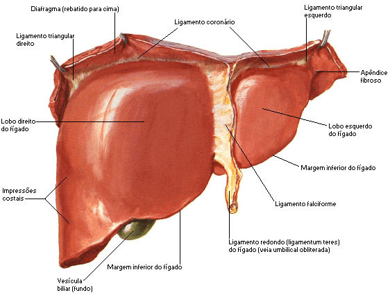
The liver has two faces: Diaphragmatic and Visceral.
The liver is divided into lobes. The Diagrammatic Face has a right lobe and a left lobe, the right being at least twice as large as the left. The division of the lobes is established by the Falciform Ligament. At the end of this ligament we find a fibrous cord resulting from the obliteration of the umbilical vein, known as the Round Ligament of the Liver .
| LIVER - DIAPHRAGMATIC FACE |
 |
| Source: NETTER, Frank H.. Atlas of Human Anatomy. 2nd edition Porto Alegre: Artmed, 2000. |
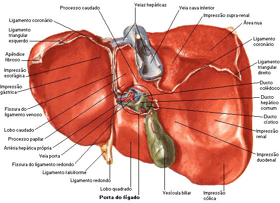
The Visceral Face is subdivided into 4 lobes (right, left, square and caudate) due to the presence of depressions in its central area, which together form an “H”, with 2 anteroposterior branches and a transversal that unites them. Although the right lobe is considered by many anatomists to include the square (lower) lobe and the caudate (posterior) lobe based on internal morphology, the square and caudate lobes more properly belong to the left lobe.
| LIVER - VISCERAL FACE |
 |
| Source: NETTER, Frank H.. Atlas of Human Anatomy. 2nd edition Porto Alegre: Artmed, 2000. |
Between the right lobe and the quadrate we find the gallbladder and between the right lobe and the caudate, there is a groove that houses the inferior vena cava. Between the caudate and quadrate lobes, there is a transverse slit: the portal of the liver (hepatic pedicle), through which the hepatic artery, portal vein, common hepatic duct, nerves and lymphatic vessels pass.
Liver Excretory Apparatus – It is formed by the hepatic duct, gallbladder, cystic duct and common bile duct.
The liver is a vital organ, being essential the functioning of at least 1/3 of it - in addition to the bile that is indispensable in the digestion of fats - it plays the important role of storing glucose and, to a lesser extent, iron, copper and vitamins.
The Liver's Digestive Function is to produce bile, a yellowish green secretion, to pass into the duodenum. Bile is produced in the liver and stored in the gallbladder, which releases it when fats enter the duodenum. Bile emulsifies fat and distributes it to the distal part of the intestine for digestion and absorption.
Other Liver Functions are:
- Carbohydrate metabolism;
- Lipid metabolism;
- Protein metabolism;
- Processing of drugs and hormones;
- Bilirubin excretion;
- Excretion of bile salts;
- Storage;
- Phagocytosis;
- Activation of vitamin D.
GALLbladder
The AV gallbladder (7-10 cm long) is located in the gallbladder fossa on the visceral surface of the liver. This fossa is located at the junction of the right lobe and the square lobe of the liver. The relationship of the gallbladder to the duodenum is so intimate that the upper part of the duodenum is normally stained with bile in the corpse. The gallbladder holds up to 50 ml of bile.
The cystic duct (4 cm long) connects the gallbladder to the common hepatic duct (union of the right and left hepatic duct) forming the common bile duct . The length varies from 5 to 15 cm. The common bile duct descends posterior to the upper part of the duodenum and lies on the posterior surface of the head of the pancreas. On the left side of the descending part of the duodenum, the common bile duct comes into contact with the main pancreatic duct.
| GALLbladder |
 |
| Source: NETTER, Frank H.. Atlas of Human Anatomy. 2nd edition Porto Alegre: Artmed, 2000. |
PANCREAS
The pancreas produces through an exocrine secretion the pancreatic juice that enters the duodenum through the pancreatic ducts, an endocrine secretion produces glucagon and insulin that enter the blood. The pancreas produces 1200 – 1500 ml of pancreatic juice daily.
The pancreas is flattened in the anteroposterior direction, it has an anterior and a posterior face, with an upper and lower border, and its location is posterior to the stomach.
The length varies from 12.5 to 15 cm and its weight in women is 14.95 g and in men 16.08 g.
The pancreas is divided into Head (located in the curve of the duodenum), Colon , Body (divided into three parts: anterior, posterior and inferior) and Tail .
| PARTS OF PANCREAS |
 |
| Source: NETTER, Frank H.. Atlas of Human Anatomy. 2nd edition Porto Alegre: Artmed, 2000. |
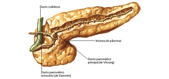
Pancreatic Duct – The main pancreatic duct begins at the tail of the pancreas and runs to its head where it curves inferiorly and is closely related to the common bile duct. The pancreatic duct joins the common bile duct (liver and gallbladder) and enters the duodenum as a common duct called the hepatopancreatic ampulla.
| CHOLEDOCH AND PANCREATIC DUCT |
 |
| Source: NETTER, Frank H.. Atlas of Human Anatomy. 2nd edition Porto Alegre: Artmed, 2000. |
The pancreas has the following functions:
- Dissolve carbohydrate (pancreatic amylase);
- Dissolve proteins (trypsin, chymotrypsin, carboxypeptidase and elastase);
- Dissolve triglycerides in adults (pancreatic lipase);
- Dissolve nucleic acids (ribonuclease and deoxyribonuclease).
